Conditions
Chondromalacia Patella Treatment in Doha
Quick answer
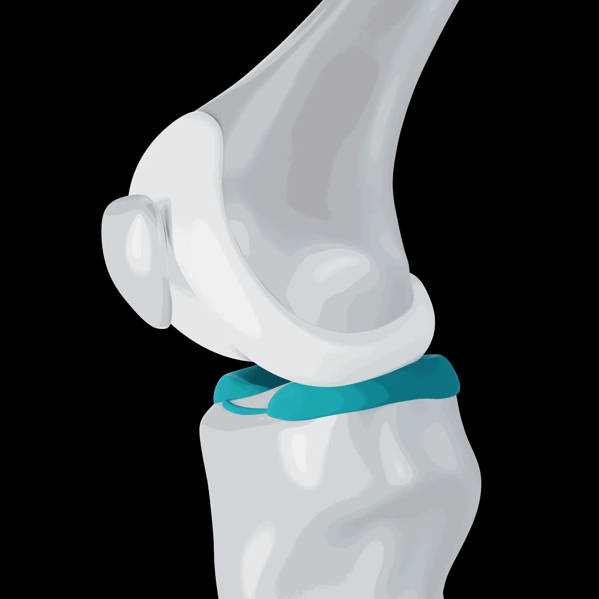
Chondromalacia patella means the cartilage on the back of the kneecap has softened or worn. It often appears on MRI reports, but the image does not always explain the pain, and most patients improve with strengthening and load management, explains Dr. Pablo Gelber, who treats kneecap problems at Aman Hospital in Doha.
What does chondromalacia patella mean?
The underside of the kneecap is covered by the thickest cartilage in the body. Chondromalacia literally means softening of cartilage. Surgeons and radiologists use it to describe changes ranging from mild softening and swelling of the surface to fissures, fraying and, in advanced cases, exposed bone.
The term is sometimes used loosely as another name for front-of-knee pain. In current practice it describes what the cartilage looks like, while patellofemoral pain describes the symptom. The two often overlap, but many people with cartilage softening on MRI have no pain at all, and many people with kneecap pain have normal cartilage.
Grades you may see on a report
- Grade 1: softening or swelling of the cartilage, with an intact surface
- Grade 2: superficial fissures or fraying, less than half the cartilage depth
- Grade 3: deeper fissures, more than half the depth
- Grade 4: full-thickness loss with bone exposed
The grade helps describe the joint, but treatment is guided far more by your symptoms, examination and goals than by the number on the report. Low-grade changes are common, especially after the age of 30.
Symptoms and causes
When chondromalacia is painful, patients describe an ache behind or around the kneecap on stairs, squatting, kneeling and after sitting, sometimes with grinding and mild swelling after activity. Causes and contributors include a previous kneecap dislocation or a direct blow such as a fall or dashboard injury, a kneecap that tracks off-centre in its groove, weakness of the thigh and hip muscles, repetitive kneeling and squatting at work or in sport, excess body weight, and age-related change. In Qatar we often see it in footballers, padel players, people who do frequent heavy squats in the gym, and in workers who kneel for long periods.
Diagnosis
Examination looks at kneecap tracking, tenderness, swelling, muscle strength and the position of the hip and foot. X-rays, including a view of the kneecap in its groove, show alignment and any arthritis. MRI shows the cartilage in detail and can detect loose fragments, bone bruising or other injuries. The key step is matching the findings to your symptoms, so that an incidental finding is not treated as the cause of every knee ache.
Non-surgical treatment
Most patients are treated without surgery. The programme is similar to that for patellofemoral pain: progressive quadriceps and hip strengthening, temporary reduction of deep squats, kneeling and hill running, low-impact cardio such as cycling or swimming, and weight management where relevant. Taping or a soft brace may give short-term relief. Anti-inflammatory medication can help during a flare. Injections, such as hyaluronic acid or platelet-rich plasma, are sometimes discussed for persistent symptoms; the evidence for them in kneecap cartilage problems is limited and mixed, and any benefit is more likely when they are combined with exercise.
Surgical options
Surgery is considered only for a minority: patients with mechanical symptoms from a loose flap of cartilage, a well-defined defect after an injury, or kneecap maltracking and instability that keeps loading the damaged area. Options include arthroscopic smoothing of unstable flaps, cartilage restoration procedures for focal defects, and realignment procedures such as tibial tubercle osteotomy or MPFL reconstruction when tracking or instability is the underlying cause. Simply smoothing cartilage for pain alone, without a mechanical problem, usually helps little.
When to see a knee specialist in Doha
Book an assessment if kneecap pain persists despite two to three months of exercise, if the knee swells repeatedly, catches or locks, if you have had a kneecap dislocation, or if an MRI report has left you worried about what the findings mean. Dr. Gelber reviews your scans with you at Aman Hospital in Doha and explains whether the cartilage findings are the likely cause of your pain.
Frequently asked questions
Can kneecap cartilage grow back?
Adult cartilage has very limited ability to heal itself. However, symptoms often improve a great deal with strengthening, even when the cartilage looks the same on a later scan. For selected focal defects, cartilage restoration surgery can help.
Is chondromalacia the same as arthritis?
Not exactly. Chondromalacia describes cartilage softening or damage, which can be limited to one area. Arthritis is a wider, usually progressive condition of the joint. Advanced grade 4 changes behind the kneecap overlap with patellofemoral arthritis.
Can I still go to the gym?
Yes, usually with adjustments. Leg presses and squats in a comfortable range, cycling, rowing and upper-body work are often fine. Deep loaded squats, jumping lunges and repeated kneeling may need to be reduced while symptoms settle.
Glossary
Patients also ask
- Can I still squat, lunge and play sport with chondromalacia patella?
- Can kneecap cartilage heal, or is chondromalacia permanent?
- Is it chondromalacia patella or runner's knee? What is the difference?
- Does chondromalacia patella need surgery?
- My kneecap grinds and hurts underneath when I go down stairs. Is it chondromalacia?
- My MRI report says chondromalacia patella grade 2 or 3. What do the grades mean?
- What is chondromalacia patella?
Related