Glossary
Long-leg alignment X-ray
Quick answer
A long-leg alignment X-ray is a single standing image of the whole leg from hip to ankle. It measures whether weight passes through the centre of the knee or is shifted inwards (bow legs) or outwards (knock knees), which matters for arthritis and surgical planning, explains Doha knee surgeon Dr. Pablo Gelber at Aman Hospital.
What to expect
You stand still on both legs against a tall X-ray plate, with kneecaps facing forward. The machine takes one long image, or several that are stitched together. It takes a few minutes and is painless. Keeping the knees straight and the feet in position matters, because rotation can change the measurements.
What it tells the surgeon
The surgeon draws a line from the centre of the hip to the centre of the ankle, called the mechanical axis. Ideally it passes close to the middle of the knee. If it falls on the inner side, that compartment carries more load, which can speed up wear and stress a repaired meniscus or cartilage.
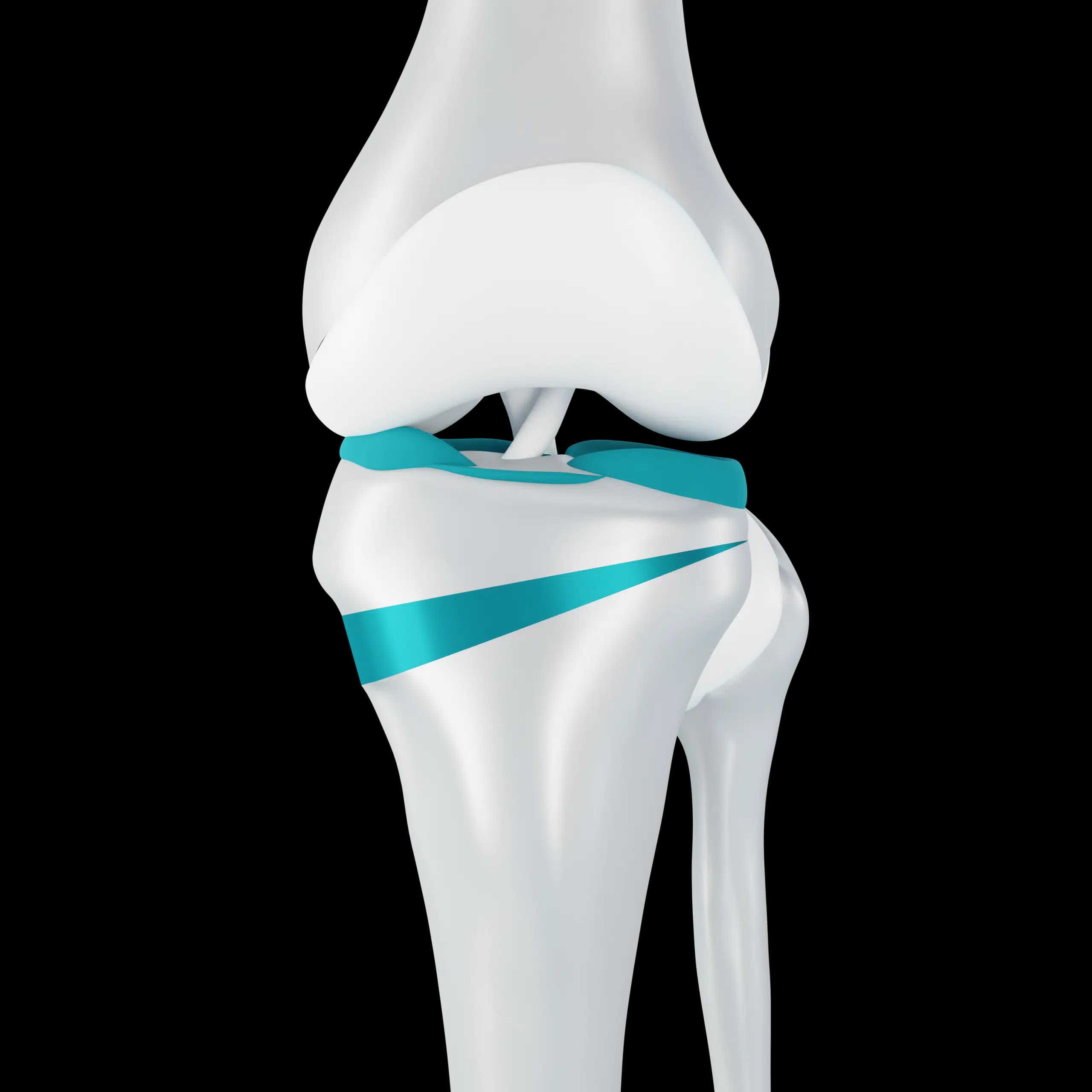
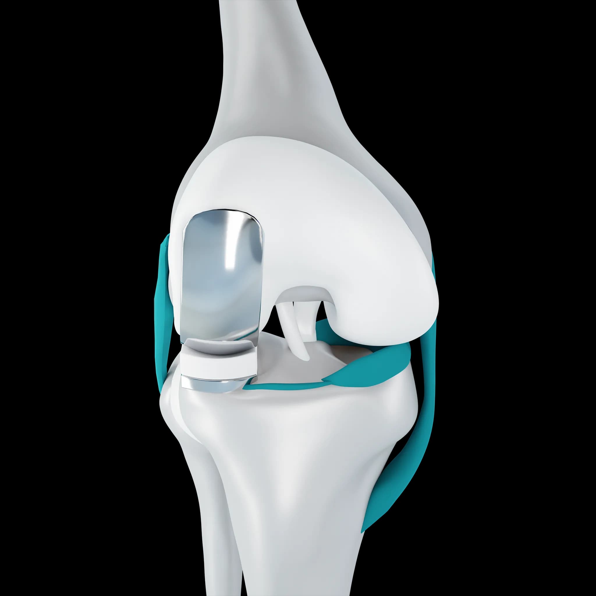
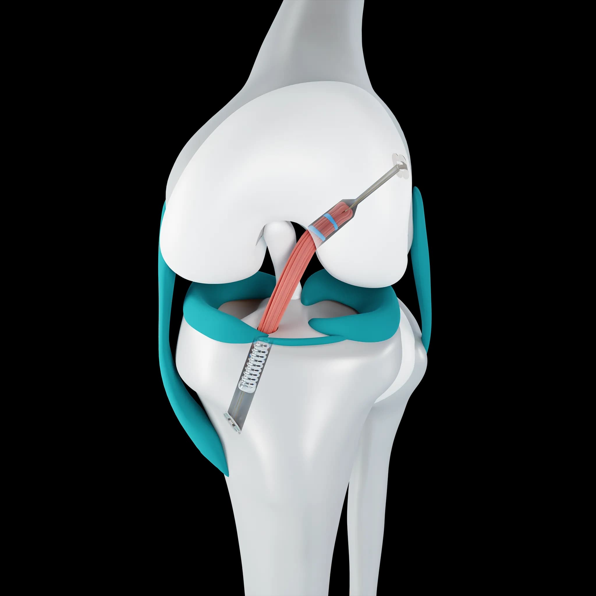
This measurement guides several decisions: whether a high tibial osteotomy could shift weight off a worn compartment, whether alignment should be corrected alongside cartilage restoration or meniscus surgery, how to plan a knee replacement, and why an ACL graft or MPFL reconstruction may be under extra strain. It is also used in teenagers with marked bow legs or knock knees.
Frequently asked questions
Why do I need a long-leg X-ray when my knee X-ray was done?
A standard knee X-ray shows only the joint. Alignment depends on the whole leg, including the hip and ankle, so a long image is needed to measure it accurately.
Can alignment be corrected?
Yes, in suitable patients. An osteotomy cuts and realigns the bone to shift load. Whether it is right for you depends on age, activity and the pattern of wear.
Related