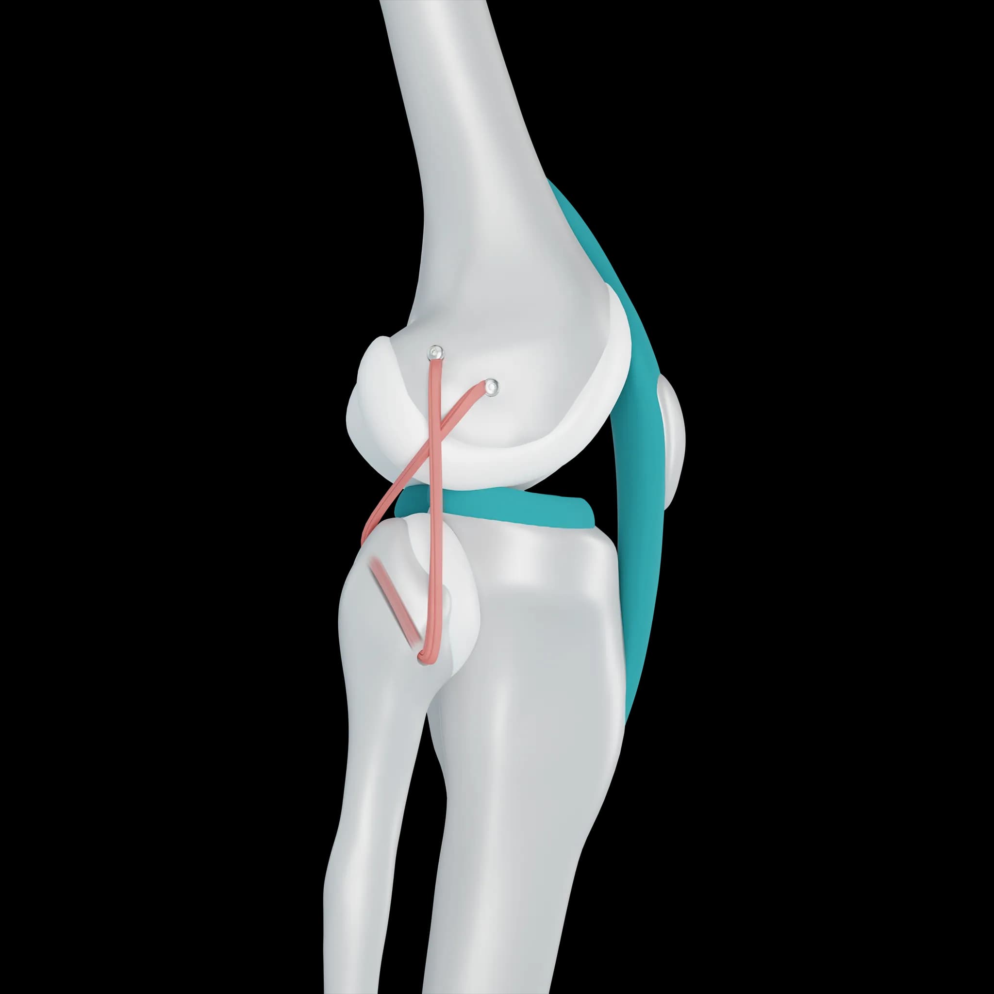
Patient question
The MRI says I have a cartilage defect in my knee. What does that mean?
 Dr. Pablo GelberOrthopaedic Knee Surgeon
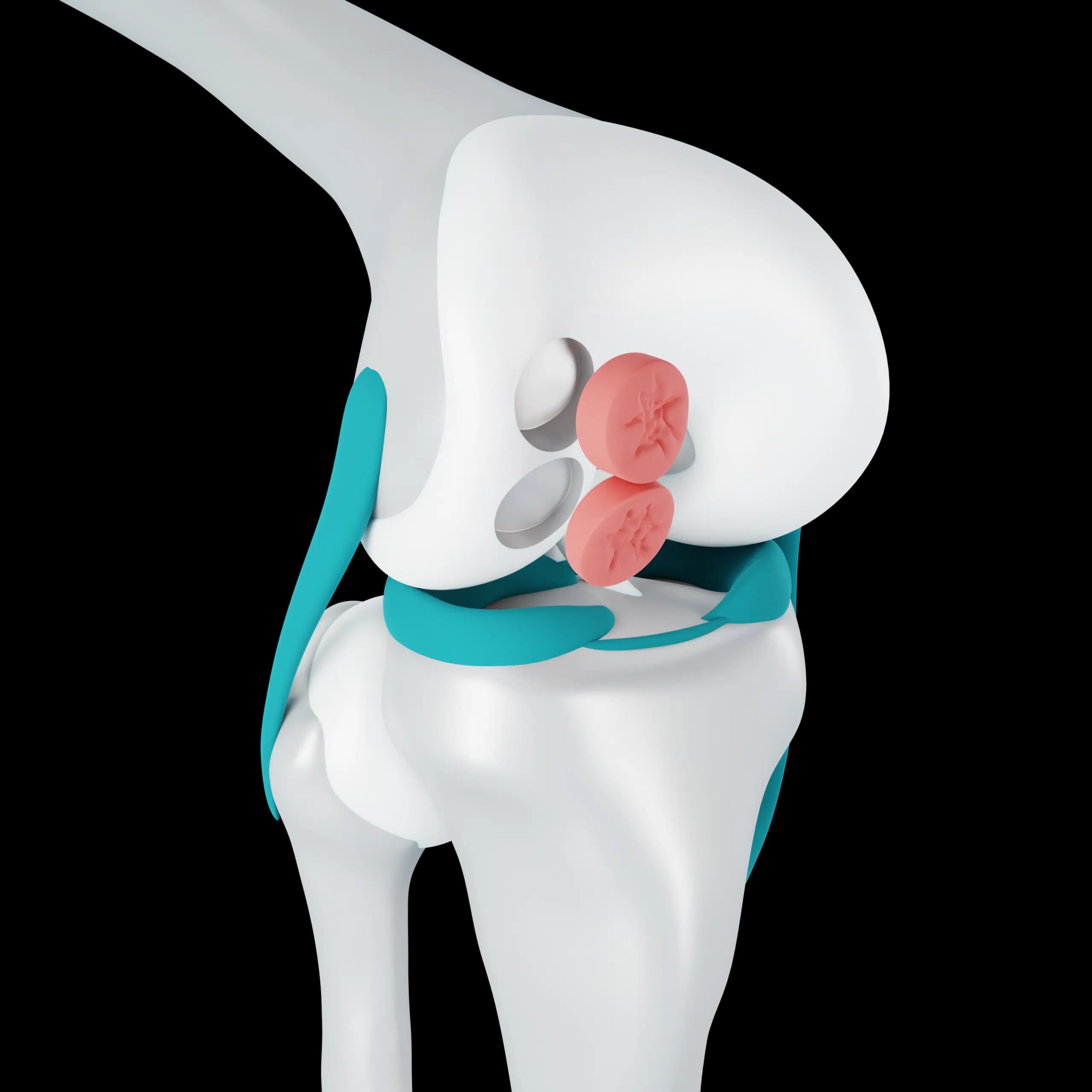
Quick answer
A cartilage defect on MRI means part of the smooth lining on the end of a bone is thinned, cracked or missing. What matters is its depth, size and location, whether the bone beneath is involved, and whether it matches your symptoms, says Dr. Pablo Gelber, knee surgeon at Aman Hospital in Doha.
Reading the words on your report
Radiologists use several terms for the same idea. Chondral defect, chondral lesion and chondropathy all describe damage to the joint cartilage. Osteochondral lesion means the bone under the cartilage is involved too.
- Partial thickness or low grade: the surface is softened or frayed but not worn through
- Full thickness or high grade: the cartilage is worn down to the bone in that spot
- Bone marrow oedema: the bone under the defect is reacting to extra load, often linked to pain
- Loose body: a fragment floating in the joint, which can cause locking
Why the location matters
The knee has three areas that bear load: the inner side, the outer side and the groove behind the kneecap. A defect on the main weight-bearing surface of the thigh bone behaves differently from one behind the kneecap, and each is treated differently. Your report usually names the area, for example medial femoral condyle or patella.
Does the MRI finding explain my pain?
Not always. Small, low-grade changes are common on MRI in adults who have no symptoms at all. A surgeon matches the scan to where you hurt, when it hurts, whether the knee swells or catches, and what the examination shows. A defect that fits the story is taken more seriously than one found by chance.
The report also needs reading alongside the meniscus, ligaments and leg alignment, because those often decide whether a cartilage defect will cope or keep getting worse.
What happens next
Bring the images, not only the written report, to your appointment. At Aman Hospital in Doha, Dr. Gelber reviews the MRI himself, examines the knee, and may request standing or long-leg X-rays to check alignment. Many small defects are treated first with physiotherapy and load management. Larger, symptomatic or unstable lesions may need a discussion about arthroscopy or cartilage restoration.
Frequently asked questions
Does a cartilage defect mean I have arthritis?
Not necessarily. A focal defect is a localised area of damage in an otherwise healthy joint. Arthritis is more widespread wear, although an untreated defect can progress over time.
Can the MRI underestimate the damage?
Yes. MRI is very useful but not perfect for cartilage, and the exact size and depth are sometimes confirmed only at arthroscopy.
Do I need a new MRI if mine was done abroad?
Often the existing scan is enough if it is recent and of good quality. A new one may be suggested if symptoms have changed or the images are hard to interpret.
Related