Patient question
My MRI says grade 2 meniscus signal. Is that a tear, and do I need surgery?
 Dr. Pablo GelberOrthopaedic Knee Surgeon
Quick answer
Grade 2 meniscus signal on MRI usually means wear or degeneration inside the meniscus that does not reach its surface, so it is generally not a true tear and rarely needs surgery. A grade 3 signal reaching the surface is a tear. Dr. Pablo Gelber, knee surgeon in Doha, always reads the scan alongside the examination.
How radiologists grade the meniscus
On MRI a healthy meniscus looks uniformly dark. Brighter areas inside it are called signal, and they are commonly graded:
- Grade 1: a small bright spot inside the meniscus, usually early wear and common with age
- Grade 2: a bright line inside the meniscus that stays within the tissue and does not reach the surface
- Grade 3: a bright line that reaches the upper or lower surface, which is the usual definition of a tear
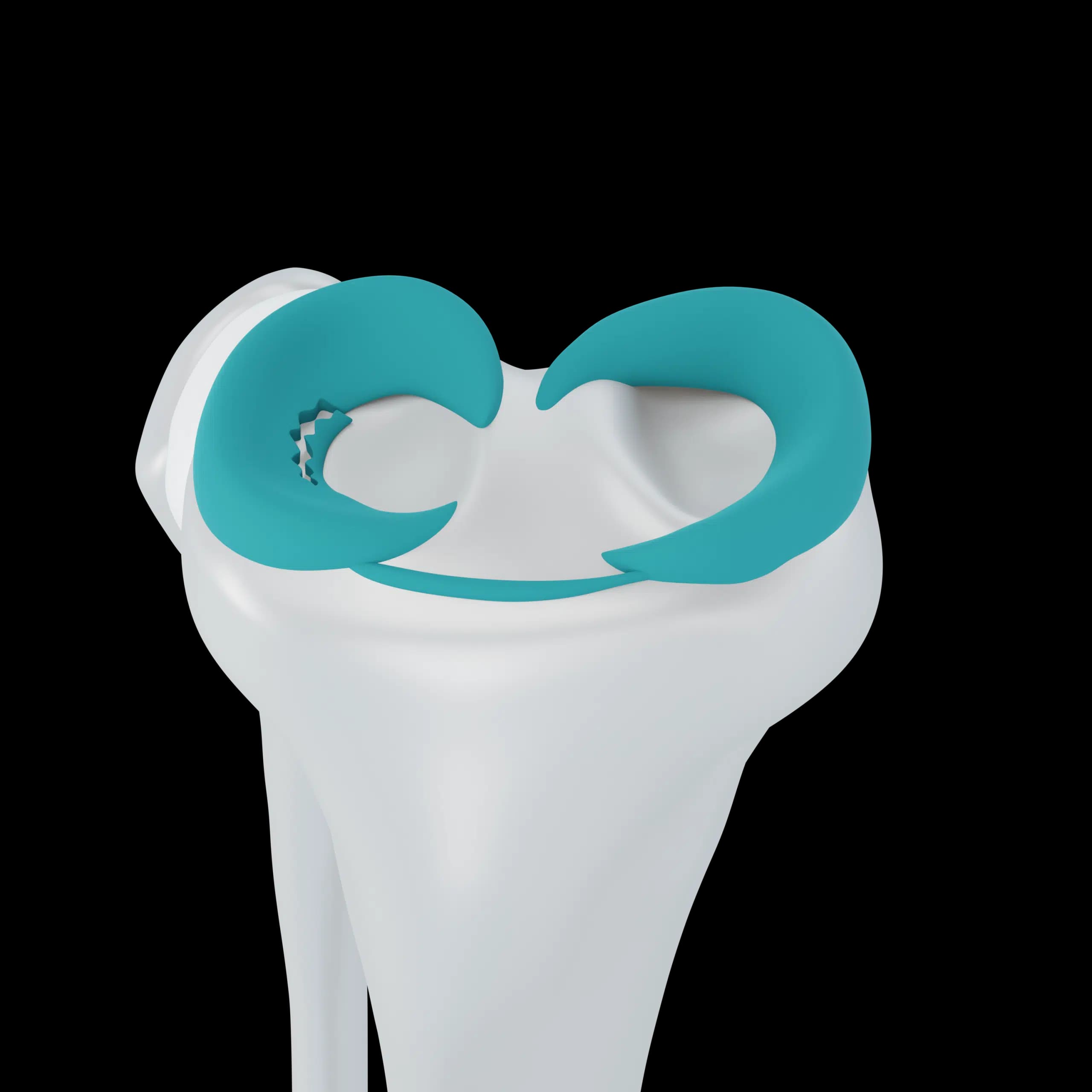
Reports may also describe the tear shape, such as horizontal, radial, vertical, flap, complex or bucket-handle, and its position, such as the posterior horn or the root.
Why the scan alone does not decide treatment
Meniscus changes are very common on MRI from middle age onwards, including in knees that cause no trouble at all. A tear on the scan therefore does not prove it is the source of your pain. Pain might come instead from early arthritis, the kneecap, a tendon, or swelling of the bone beneath the cartilage.
The opposite also happens. Occasionally a small tear is hard to see, particularly at the back of the lateral meniscus or near the root, and a surgeon's examination raises suspicion that a second look at the images confirms.
What usually happens with grade 2
Grade 2 signal is managed like a sore, ageing meniscus rather than a tear: strengthening, load management and time. It does not need to be cut out. Some grade 2 areas progress to a tear over the years, but most do not cause mechanical symptoms.
Getting your MRI reviewed in Doha
Bring the images, not only the written report, to your appointment. At Aman Hospital, Dr. Gelber looks at the scan himself and matches it to where you hurt, how the knee behaves and what he finds on examination. For tears that are suspicious of a root or bucket-handle pattern, earlier treatment can matter, and this is explained clearly.
Frequently asked questions
My report says complex degenerative tear. Is that serious?
It describes a worn meniscus with several tear lines, common after about 45. It is usually treated with physiotherapy first. It becomes more significant if the knee locks or if arthritis is also present.
Should I repeat the MRI to see whether the tear has healed?
Usually not. Treatment is guided by how the knee feels and functions. A repeat scan is reserved for new symptoms or when surgery is being planned.
What is bone marrow oedema on my report?
It is fluid within the bone, which can come from a bruise, overload or, in older knees, a stress reaction under a root tear. It should be interpreted by a knee surgeon together with your symptoms.
Related