Patient question
Do I need an MRI after my kneecap dislocated, if the X-ray was normal?
 Dr. Pablo GelberOrthopaedic Knee Surgeon
Quick answer
Often, yes. X-rays show bone but can miss cartilage that was sheared off when the kneecap slipped, as well as the ligament tear itself. After a first dislocation, especially with marked swelling, an MRI is commonly recommended to plan treatment, explains Dr. Pablo Gelber, knee surgeon at Aman Hospital in Doha, Qatar.
What an X-ray can and cannot see
X-rays are quick and useful straight after the injury. They confirm the kneecap is back in place, show larger bone fragments and give a first look at the groove and kneecap height.
They cannot show cartilage, which is invisible on plain films, or ligaments. A cartilage flake knocked off the back of the kneecap or the edge of the thigh bone may therefore be present even when the X-ray report says normal.
What an MRI adds
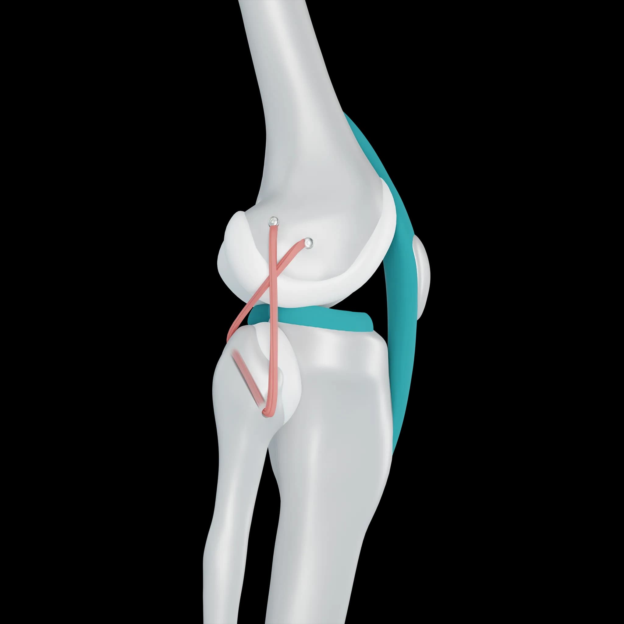
- Whether the medial patellofemoral ligament (MPFL) is torn, and where along its length
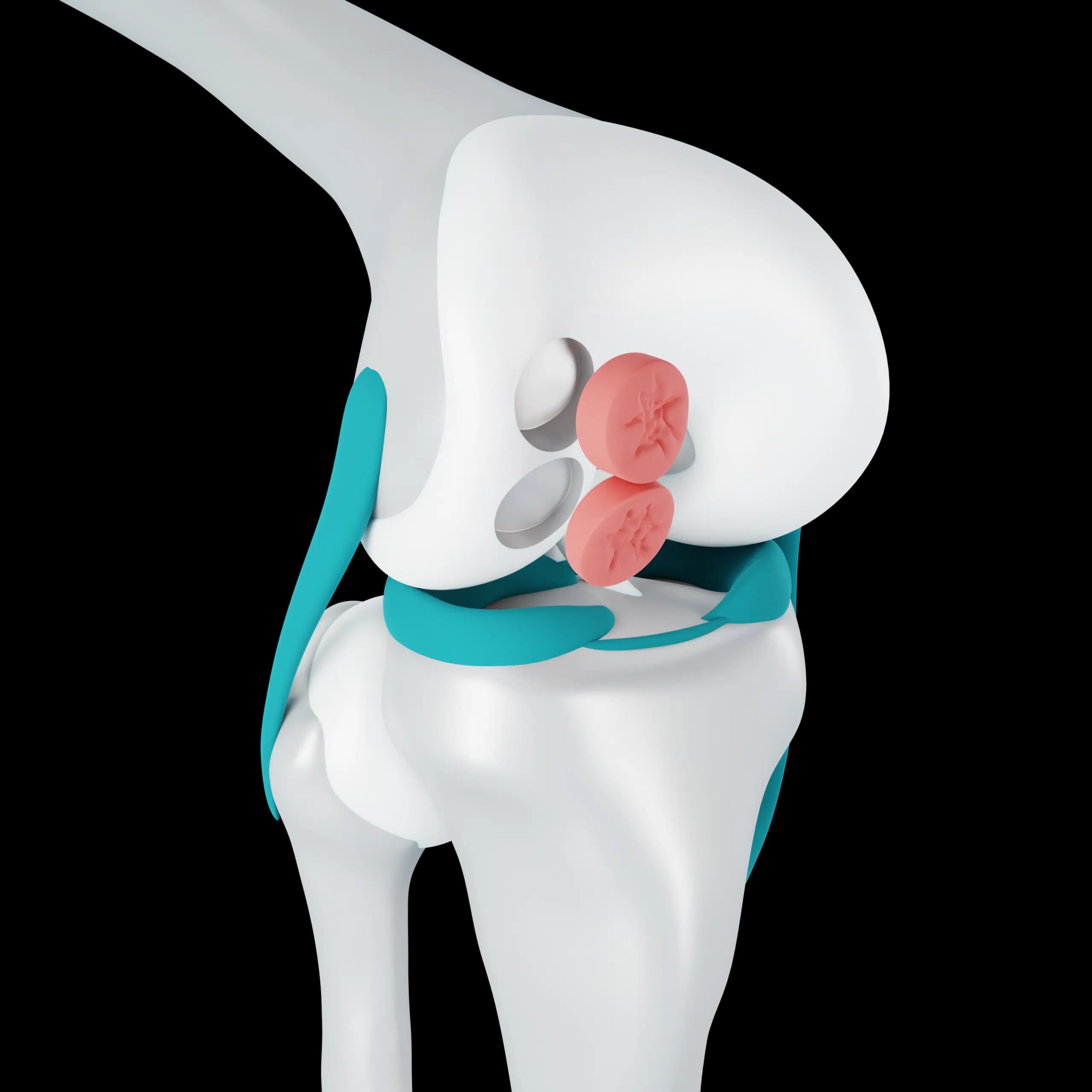
- Cartilage injury on the back of the kneecap or the outer edge of the thigh bone
- Loose fragments of cartilage or bone inside the joint
- Bone bruising, which confirms a dislocation happened even if it reduced by itself
- Measurements of the groove depth, kneecap height and tendon position, which help predict the risk of recurrence
When it changes the plan
If the MRI shows a sizeable loose fragment, early surgery is often advised to fix it back or remove it, because a loose piece can catch, lock the knee and wear the joint. If there is no fragment, the result usually supports starting physiotherapy, and the measurements help decide how cautious to be with sport.
For some patients with mild swelling and a clear examination, the surgeon may judge that an MRI can wait. That decision depends on the examination.
Arranging imaging in Doha
At Aman Hospital, Dr. Gelber reviews any emergency X-rays you bring, examines the knee and arranges an MRI when it will help. He then goes through the images with you, pointing out the ligament, the cartilage and the groove, so the reason for the recommended treatment is clear.
Frequently asked questions
How soon after the injury should the MRI be done?
Usually within the first couple of weeks, so that a loose fragment can be dealt with promptly if one is found. Your surgeon will advise on timing.
Will I need a CT scan as well?
Not usually. A CT is sometimes added before surgery to measure bone rotation or the tendon position in more detail.
Related