Glossary
Subchondral bone
Quick answer
Subchondral bone is the layer of bone directly beneath the articular cartilage. It supports the cartilage, absorbs load and, unlike cartilage, has nerves, so it is often where knee pain from arthritis or injury actually comes from, notes Doha-based knee surgeon Dr. Pablo Gelber of Aman Hospital.
What it is
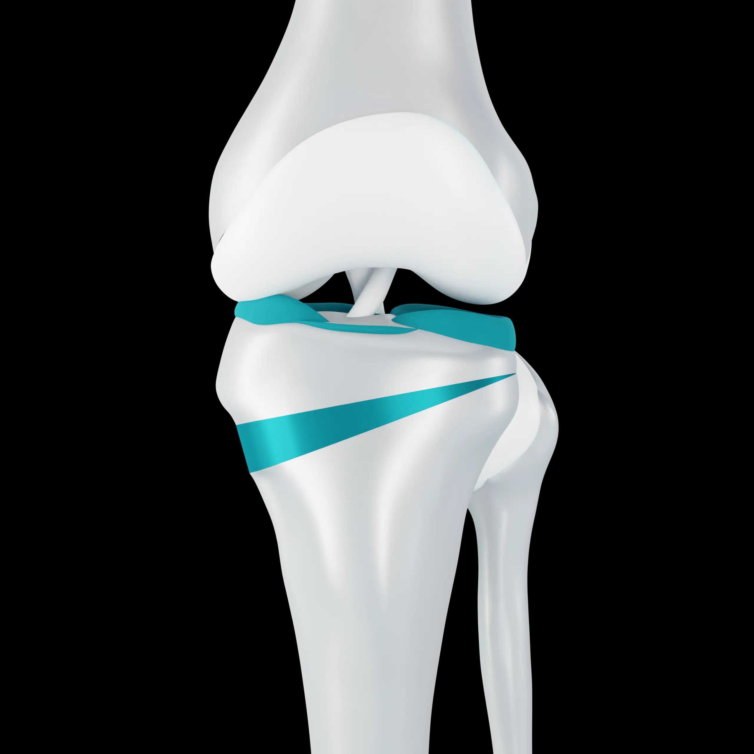
Under the cartilage lies a thin, dense plate of bone, and beneath that a spongy network of bone and marrow. Together they act as a shock absorber for the cartilage above. The cartilage and the subchondral bone behave as one unit: when one is damaged, the other is usually affected.
Why it matters for patients
MRI and X-ray reports often mention this layer. Bone marrow oedema, sometimes described to patients as water on the bone, is increased fluid in the bone and appears after impact injuries and bone bruises, with stress reactions, and in active arthritis. Subchondral sclerosis means the bone has become denser, and subchondral cysts are small fluid-filled holes; both are common signs of osteoarthritis on X-ray.
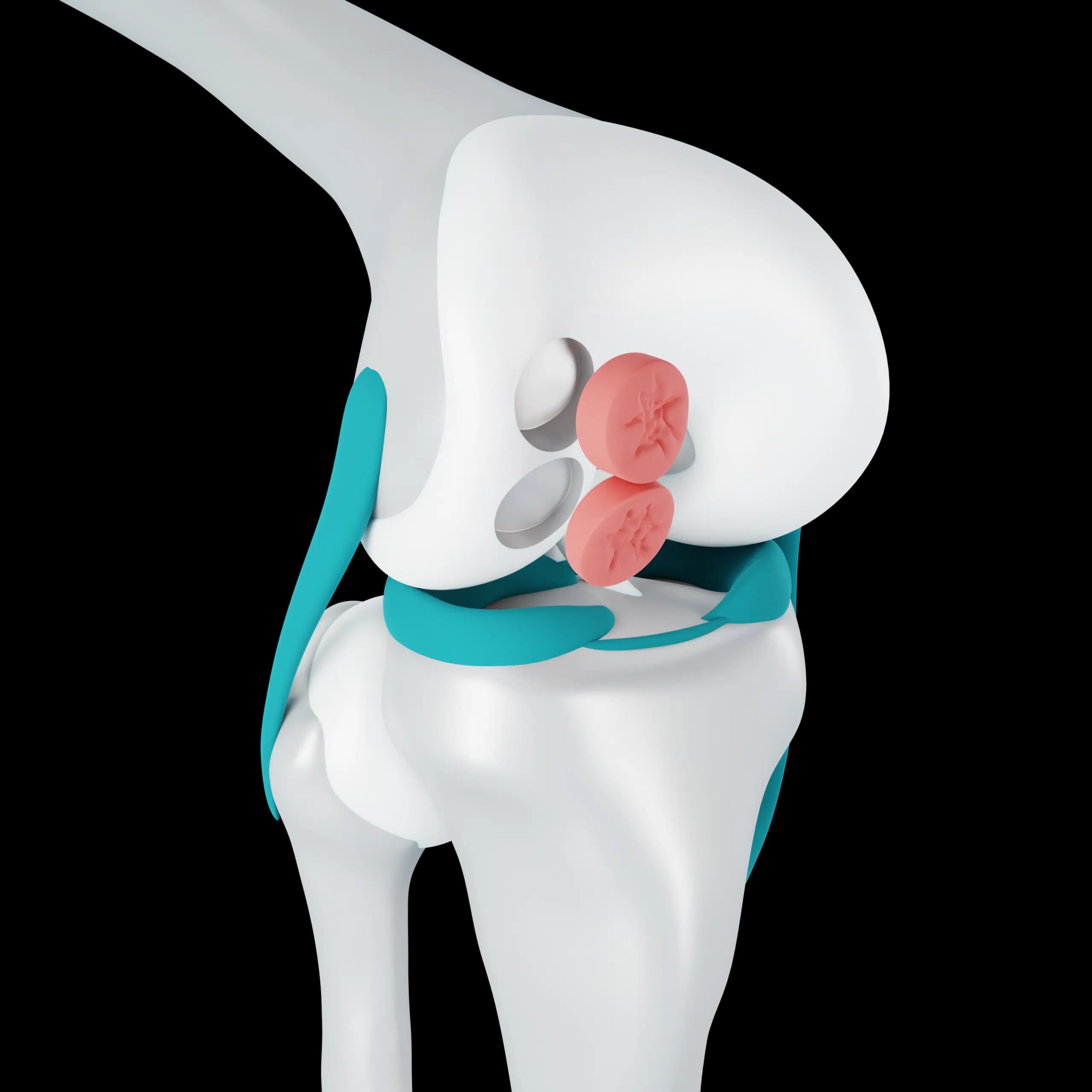
In osteochondritis dissecans, a piece of this bone and its cartilage loosens. In older adults, a sudden painful area of bone collapse, called a subchondral insufficiency fracture, can follow minor strain, sometimes alongside a meniscal root tear. These terms sound alarming but are usually explained and managed with load modification, physiotherapy and time, with surgery reserved for specific situations.
Frequently asked questions
My MRI says bone marrow oedema. Is it serious?
It is a common finding that means irritation or bruising of the bone. Its significance depends on where it is, what caused it and your symptoms, which your surgeon will explain.
Why does arthritis pain come from bone if cartilage is the problem?
Cartilage has no nerves. As it thins, more load reaches the subchondral bone, which does have nerves, and that bone stress is a major source of arthritis pain.
Related