Glossary
Femur (thigh bone)
Quick answer
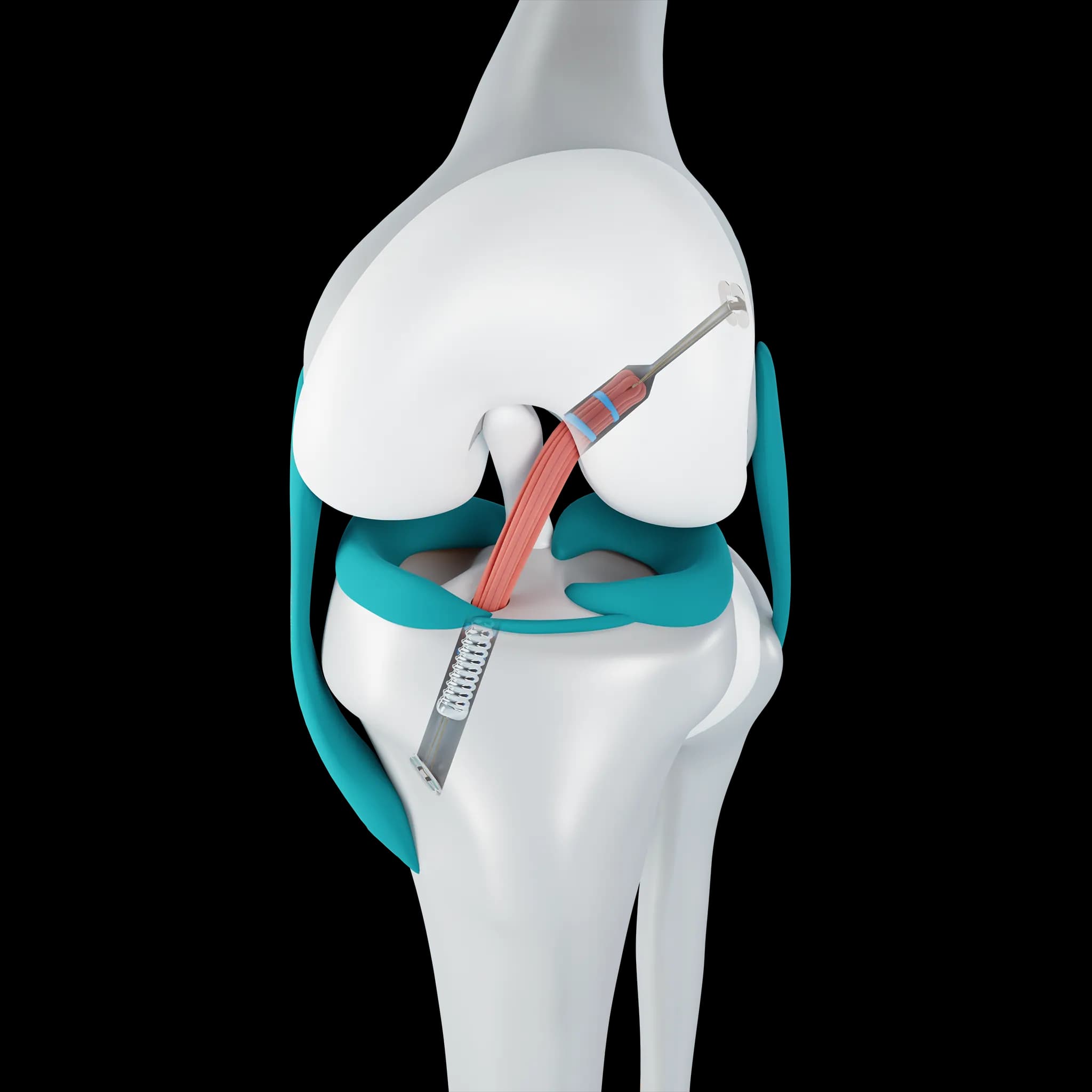
The femur is the thigh bone, the longest and strongest bone in the body. Its lower end widens into two rounded condyles that form the top half of the knee joint and the groove the kneecap runs in, explains Dr. Pablo Gelber, knee surgeon at Aman Hospital in Doha.
What it is
The femur runs from the hip to the knee. At the knee, its lower end (the distal femur) flares into the medial and lateral femoral condyles, which roll and glide on the top of the tibia. Between them at the front lies the trochlea, the groove the kneecap slides in. At the back, a notch between the condyles houses the cruciate ligaments.
The joint surfaces of the femur are covered with smooth articular cartilage, and the bone just beneath it, the subchondral bone, supports that cartilage.
Why it matters for patients
Many knee problems start on the femoral side of the joint. Cartilage wear on the condyles is a common part of knee osteoarthritis. In young people, osteochondritis dissecans usually affects a femoral condyle. Runners and military recruits can develop stress fractures in the lower femur, and twisting injuries often leave bone bruises there. The shape of the femur, including its rotation and the depth of the trochlea, also affects how stable the kneecap is.
How it is examined
Standing X-rays show the femoral bone, its alignment with the tibia and the joint space. An MRI shows cartilage, bone bruising and early stress injuries that X-rays can miss. A CT scan is sometimes used to measure rotation or plan surgery.
Frequently asked questions
Can knee pain come from the femur itself?
Yes. Cartilage damage on the femoral condyles, bone bruises, stress fractures and osteochondritis dissecans all involve the femur and are felt as knee pain.
Does knee replacement remove the femur?
No. A knee replacement only resurfaces the worn ends of the bones. A thin layer of the lower femur is shaped to hold a metal component; the rest of the bone stays as it is.
Related